
Could the Resonant Recognition Model explain how Frequency Specific Microcurrent Works?
- ResonantEquus

- Feb 27
- 7 min read
UNDERSTANDING THE RESONANT RECOGNITION MODEL (RRM) MAY BE THE SCIENTIFIC KEY TO HOW FREQENCY SPECIFIC MICROCURRENT (FSM) ACTUALLY WORKS


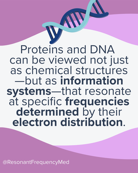


The Resonant Recognition Model (RRM) is a theoretical scientific framework proposing that biomolecules recognize and interact with each other through specific electromagnetic resonance patterns encoded in their primary sequences, rather than only through lock‑and‑key structural complementarity.
The Core idea of Cosic's Resonant Recognition Model (RRM)
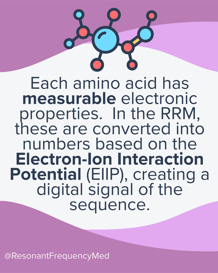
Each protein or DNA sequence is converted into a numerical signal by assigning every amino acid (or nucleotide) a value related to delocalized electron energy or similar physical property. Performing spectral analysis (a Fourier transformation) on that signal reveals that proteins sharing a biological function exhibit a common dominant frequency – their “resonant signature” – which is proposed to underlie selective recognition and interaction. RRM uses the spectra as “functional fingerprints” and looks for shared or complementary dominant frequencies between proteins to infer potential interactions. Scroll down to see a chart of the numerical value (EIIP) of each of the 20 amino acids.
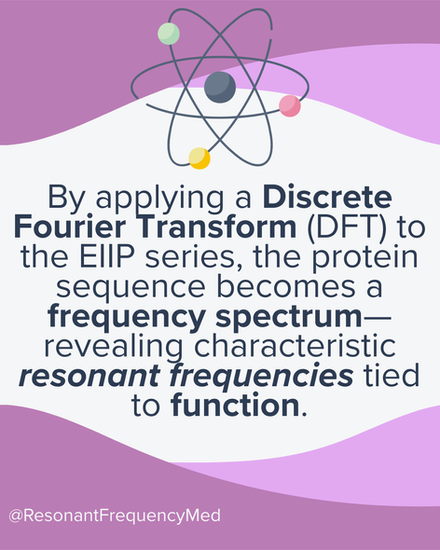
Fourier Transform
A Fourier transform is a mathematical operation that takes a signal and expresses it in terms of its frequency content instead of its original form in time or space. In more concrete terms, a signal (a curve, waveform, or sequence) can be represented as a sum of sine and cosine waves of different frequencies and amplitudes. The Fourier transform tells you “how much” of each frequency is present in the original signal; its output is called the frequency‑domain representation.
RRM Mechanism and Predictions
The Resonant Recognition Model assumes macromolecules interact via long‑range, weak electromagnetic fields at specific frequencies corresponding to these dominant spectral components. Consider reading Pollack's Charged if this topic interests you further!
Functionally related proteins (such as receptors and ligands, transcription factors and DNA sites) are predicted to share or complement characteristic RRM frequencies and wavelengths, providing a quantitative criterion for “match” or recognition. In other words, within a group of proteins with the same biological function (e.g., a family of growth factors), RRM identifies a characteristic peak frequency that is common to that functional group.
The model can highlight “hot spots” in protein sequences—residues that are not necessarily catalytic but cluster in 3D space around active sites and are proposed to shape the local structure and field environment required for binding and activity.
Applications in sequence analysis and design
RRM has been used to identify conserved, functionally important regions in protein families, based on their contribution to the characteristic resonant frequency rather than classic sequence homology alone. It has also been applied to rationally design bioactive peptides by choosing sequences that reproduce the RRM frequency of a target protein function (for example, agonist‑ or antagonist‑like activity).
Links to biophoton emission and external fields
Later work extends RRM to suggest that ultra‑weak photon emissions from cells (biophotons) occur at wavelengths matching RRM‑predicted frequencies for specific proteins or pathways, implying that functional states may be tracked or influenced optically. Some studies claim that applying light at wavelengths corresponding to RRM frequencies can modulate cellular processes (e.g., growth, signaling), supporting the idea that these resonant frequencies are not just mathematical artifacts but may be biophysically actionable.
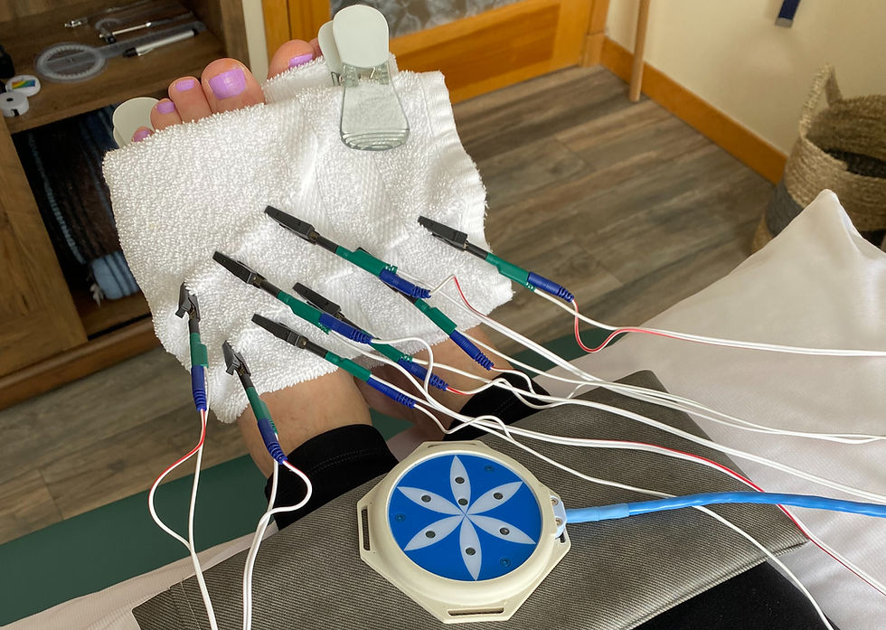
Expanding RRM into Frequency Specific Microcurrent
If this modulation of cellular processes mentioned above is in fact possible, then inputs of specific frequencies at micro-amperage levels, which is what we do in FSM, may also produce biophysically actionable results explained by the RRM.
Resonance as Frequency Information
RRM and FSM both assume that frequency is not just a carrier but part of the information itself. In FSM, we describe frequencies as instructions (channel A) and target tissues (channel B). Pairing the correct instruction or pathogen frequency with the correct target tissue frequency elicits significant and specific changes.
In RRM, a protein’s primary sequence is mapped to EIIP values and passed through a Fourier transform to yield a characteristic “functional frequency” (or band) associated with that protein’s activity and interaction partners; whereas, in FSM, we are selecting external frequencies (in Hz) that appear to “match” or neutralize specific tissues and conditions, as if each tissue/condition has its own characteristic resonance. At the conceptual level, both frameworks say: “biological specificity can be expressed in the frequency domain.” RRM does this mathematically for sequence–structure–function; FSM does it clinically for tissue–condition–response.
Proteins as frequency‑selective elements
In RRM, each protein (or family of proteins) has dominant resonant frequencies related to its delocalized electron structure and thus to its functional state and interaction patterns. Within the cell, proteins, membranes, channels, and cytoskeletal structures act like frequency‑selective elements: they respond preferentially to fields oscillating near their characteristic RRM frequencies (or harmonics). Pathologic states (inflammation, fibrosis, neuropathic sensitization) correspond to altered distributions of active proteins and conformations, which would change the “frequency landscape” in that tissue.
FSM applies low‑intensity currents at specific macroscopic frequencies (instruction frequency, tissue frequency). If some of those frequencies (or their harmonics / beat frequencies) overlap with RRM‑like resonant bands for key proteins or protein complexes in that tissue, they could bias conformational equilibria, binding probabilities, or signaling cascades, nudging the system toward a less inflamed, less sensitized, more regenerative state.
In other words: RRM says “proteins have preferred frequencies;” FSM says “tissues and conditions respond to preferred frequencies.” If conditions/in/tissues are just organized ensembles of proteins and extracellular matrix, these statements could be two scales of the same resonance principle.
Dual-channel FSM as interacting spectral targets
We can theoretically interpret FSM through an RRM‑like lens: The target tissue frequency (channel B) corresponds to the dominant resonances of the structural and functional proteins defining that tissue (e.g., neural membrane channels, cytoskeletal proteins, ECM that are described in RRM). The pathogen or instruction frequency (channel A) corresponds to the resonances of proteins and pathways that encode or regulate that state: inflammatory mediators, fibrosis‑related enzymes, ion channels, etc.
When we run dual-channel frequencies, we influence the system at a combination of frequencies that could selectively address both “what it is” (tissue identity) and “what it’s doing now” (pathologic state). If those externally applied frequencies overlap with, or modulate toward, healthier RRM‑type frequencies for the tissue’s proteins, we could see changes in:
channel gating and membrane potential
intracellular calcium handling
kinase/phosphatase activity
transcriptional programs
which matches the clinical picture of FSM (changes in pain, tone, inflammation, tissue texture, ANS balance, and more).
The good news here is that we have ways to map or measure (namely: ultrasound, labs, imaging studies, cytokine levels, AROM measures, and more) that can be the subjects of more formalized research as to how FSM works.
Why discrete “tissue” and “condition” frequencies could exist
RRM provides a plausible explanation for why discrete frequencies might be empirically discoverable in the clinic:
Different tissues express distinct protein repertoires and architectures → different aggregate resonance spectra.
Different conditions (inflammation, ischemia, fibrosis, central sensitization) shift which proteins are up‑regulated, how they’re modified (phosphorylation, conformation), and how they cluster → different emergent resonances.
When FSM pioneering clinicians “discover” a tissue or condition frequency empirically, RRM might say we're indirectly mapping the dominant spectral feature of the protein ensemble that defines that tissue, or the altered spectral feature of the pathologic protein network in that condition.
The fact that many FSM frequencies are stable and reproducible across practitioners and patients could be viewed as evidence that those protein ensembles have relatively stable resonant properties at the macroscopic scale.
How to approach this information as an FSM clinician, or as a skeptical patient
RRM is a hypothesis‑driven model that offers a unifying way to relate sequence, structure, function, and electromagnetic interactions, and it has inspired exploratory work in peptide design, photobiomodulation, quantum applications and bioelectromagnetics. The RRM, like FSM, remains outside mainstream biochemistry; evidence is mixed and largely preclinical or theoretical, so it should be viewed as a conceptual framework and potential generator of testable predictions, rather than established physiology at this stage.
However, the RRM may provide a very valuable way to explore the mechanism behind frequency specific microcurrent, in particular, as it describes that:
Proteins and protein networks have preferred electromagnetic resonance frequencies tied to their sequence and structure (as modeled by RRM).
Pathologic states shift which resonant modes dominate at the tissue level.
FSM applies microampere currents at specific frequencies that appear to couple to these resonant modes, selectively modulating the activity of proteins and pathways that define a tissue and its pathologic condition.
The clinical effects we see (changes in pain, inflammation, tone, tissue pliability, ANS function) may emerge from subtle, frequency‑specific changes in protein conformation, binding, and signaling—essentially “retuning” the tissue’s resonance profile toward a healthier state.
Are you a Frequency Specific Practitioner Interested in Exploring the Mechanisms?
Join our group of curious, open-minded, dedicated clinician-researchers as we forge a pathway to validating the incredible clinical findings we encounter in the realm of Frequency Medicine.
Who is I. Cosic and what is she up to now?
Ira (Ivanica) Cosic, often cited as I. Cosic, is a biophysicist and electrical engineer best known for developing the Resonant Recognition Model. Her work has been influential in biophysics, bioinformatics, and complementary‑medicine circles, particularly for those exploring how frequency‑based approaches might interface with biological systems.
Cosic trained and worked in Australia and later Europe, and throughout her career she published widely on RRM, protein–ligand recognition, and the use of signal‑processing methods to derive “functional frequencies” for receptors, enzymes, and other macromolecules. Her later work extended RRM ideas into bioactive peptide design, photobiomodulation‑like predictions, and exploratory uses in viral and disease‑strain differentiation, laying conceptual groundwork that some clinicians now compare with frequency‑specific microcurrent (FSM) and bioelectromagnetic therapies.
Throughout her career she has been recognized as a thoughtful, technically rigorous researcher who bridged engineering and life‑science perspectives, leaving a legacy that continues to inspire interest in the role of electromagnetic and frequency‑based phenomena in biology.
Irukayama “Iva” Cosic appears to be retired, and a direct RRM pipeline is no longer being actively extended by an academic group. However, the core idea that biomolecules or ensembles can be “addressed” or modulated by specific frequencies is being explored in several modern quantum‑biology, biophotonics, and bioelectromagnetic contexts that we may be able to draw on when exploring the connections between RRM and FSM.
RESOURCES
Cosic I. Macromolecular bioactivity: is it resonant interaction between macromolecules? IEEE Trans Biomed Eng. 1994;41(12):1101-1114.
Cosic I. The Resonant Recognition Model of macromolecular bioactivity. In: Pandalai SG, ed. Recent Research Developments in Biophysics. Vol 1. Trivandrum, India: Research Signpost; 2002:13-30.
Cosic I. Resonant recognition model and protein topography. Eur J Biochem. 1991;198(1):1-13.
Cosic I, Vojisavljevic V, Pavlovic M. The Resonant Recognition Model (RRM) predicts amino acid residues in highly conserved regions of protein sequences. J Mol Graph Model. 2001;19(3-4):248-258.
Pirogova E, Istivan T, Cosic I. Bioactive peptide design using the resonant recognition model. BMC Bioinformatics. 2007;8:353.
Cosic I, Cosic D, Lazar K. Cosic’s Resonance Recognition Model for protein sequences and photon emission differentiates lethal and non-lethal Ebola strains: implications for treatment. Open J Biophys. 2015;5(4):1-13.
Dotta BT, Murugan NJ, Karbowski LM, Lafrenie RM, Persinger MA. Shifting the wavelengths of ultraweak photon emissions from dying melanoma cells: their chemical enhancement and blocking are predicted by Cosic’s RRM. Naturwissenschaften. 2014;101(2):87-94.


Comments